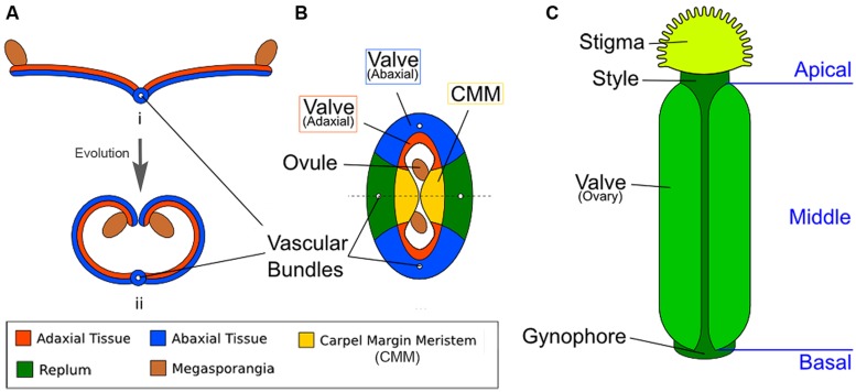
FIGURE 1.
Diagrams illustrating the homology between modern carpels and ancestral leaves. (A) Hypothetical evolution of a single plicate carpel based on Scagel (1965). (i) A cross section of an ancestral plant’s spore-bearing leaf (sporophyll), showing megasporangia at the leaf edge. (ii) Over evolutionary time, inward curling of a megasporangia-bearing leaf and subsequent fusion at the leaf margin led to a one-chamber ovary with two rows of megasporangia on the interior (adaxial side). The actual evolutionary path is more complicated and not fully settled. (B) The cross section view of the Arabidopsis gynoecium, consisting of two fused carpels enclosing two locules. Note the vascular bundles. Although there are four rows of ovules, only two ovules are visible in the cross-section since the rows alternate within each locule. (C) A diagram of the Arabidopsis gynoecium, showing that it consists of three regions along the basal-to-apical axis. The basal section consists of a short stalk, the gynophore, the middle section is the ovary, and the apex consists of style and stigma.

