

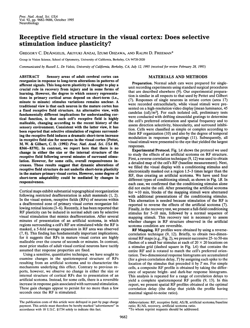
Abstract
Sensory areas of adult cerebral cortex can reorganize in response to long-term alterations in patterns of afferent signals. This long-term plasticity is thought to play a crucial role in recovery from injury and in some forms of learning. However, the degree to which sensory representations in primary cortical areas depend on short-term (i.e., minute to minute) stimulus variations remains unclear. A traditional view is that each neuron in the mature cortex has a fixed receptive field structure. An alternative view, with fundamentally different implications for understanding cortical function, is that each cell's receptive field is highly malleable, changing according to the recent history of the sensory environment. Consistent with the latter view, it has been reported that selective stimulation of regions surrounding the receptive field induces a dramatic short-term increase in receptive field size for neurons in the visual cortex [Pettet, M. W. & Gilbert, C. D. (1992) Proc. Natl. Acad. Sci. USA 89, 8366-8370]. In contrast, we report here that there is no change in either the size or the internal structure of the receptive field following several minutes of surround stimulation. However, for some cells, overall responsiveness increases. These results suggest that dynamic alterations of receptive field structure do not underlie short-term plasticity in the mature primary visual cortex. However, some degree of short-term adaptability could be mediated by changes in responsiveness.
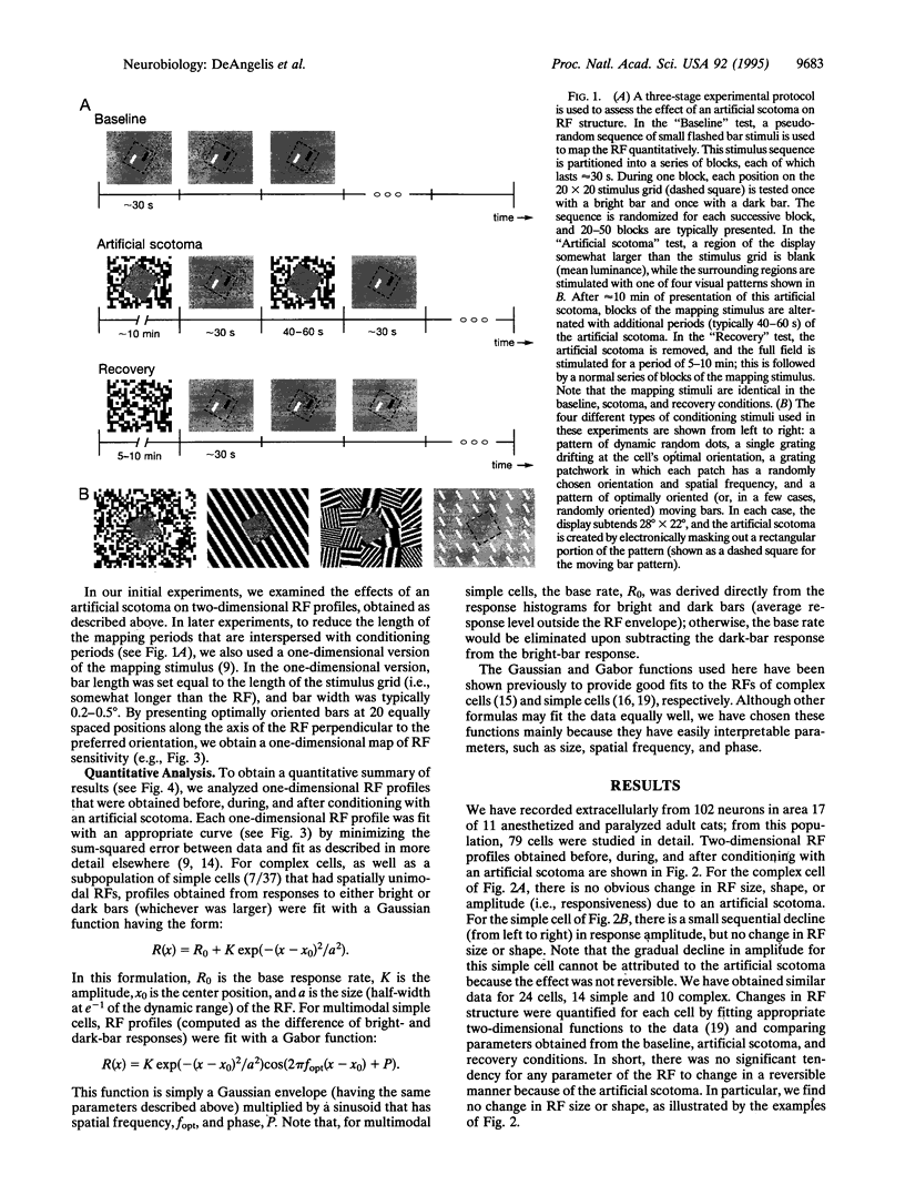
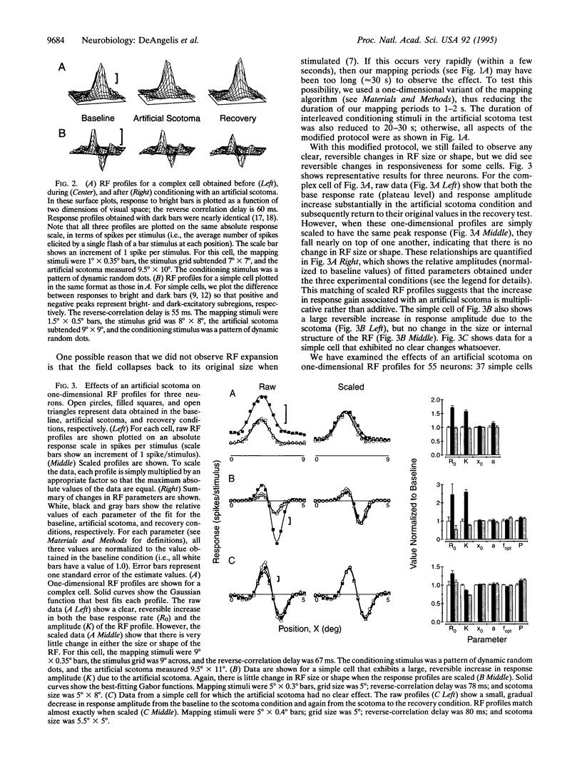
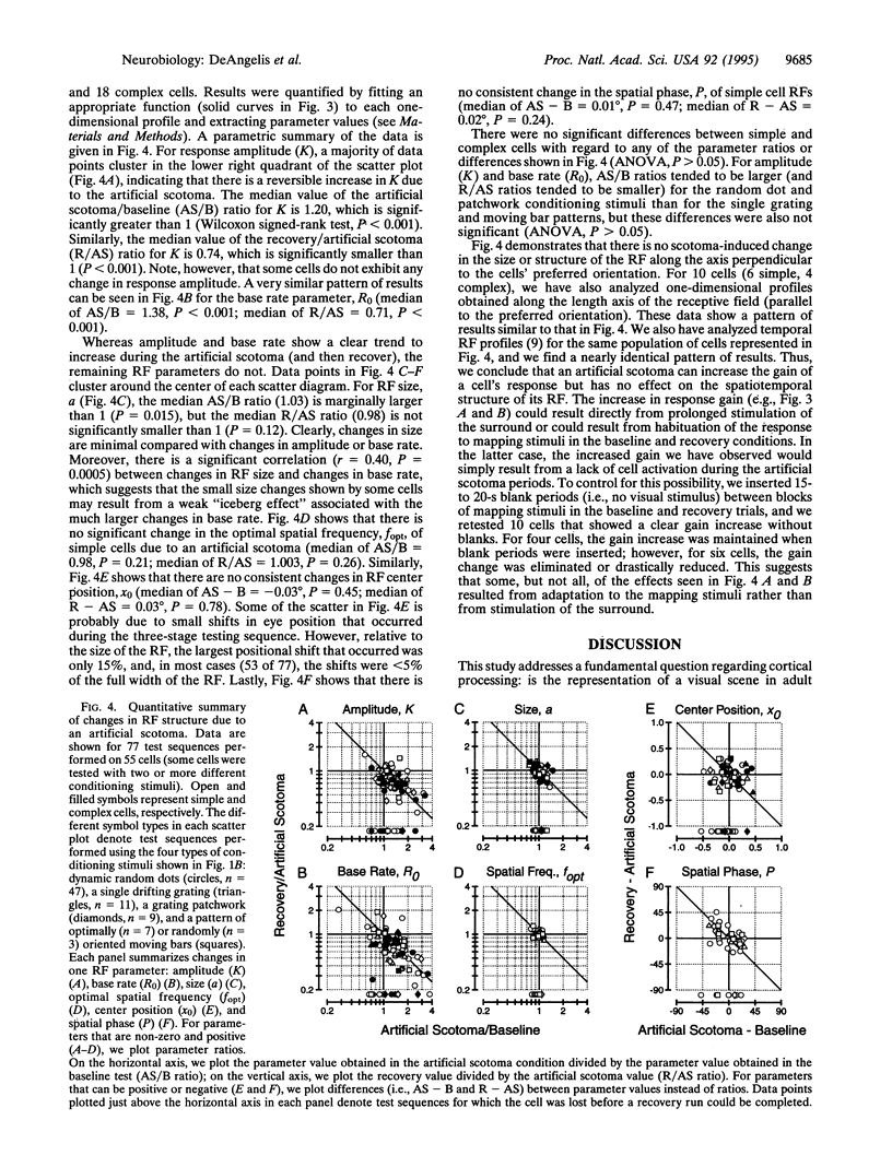
Full text
PDF
Images in this article
Selected References
These references are in PubMed. This may not be the complete list of references from this article.
- Baker C. L., Jr, Cynader M. S. Spatial receptive-field properties of direction-selective neurons in cat striate cortex. J Neurophysiol. 1986 Jun;55(6):1136–1152. doi: 10.1152/jn.1986.55.6.1136. [DOI] [PubMed] [Google Scholar]
- Barlow H. B. Single units and sensation: a neuron doctrine for perceptual psychology? Perception. 1972;1(4):371–394. doi: 10.1068/p010371. [DOI] [PubMed] [Google Scholar]
- Campbell F. W., Cleland B. G., Cooper G. F., Enroth-Cugell C. The angular selectivity of visual cortical cells to moving gratings. J Physiol. 1968 Sep;198(1):237–250. doi: 10.1113/jphysiol.1968.sp008604. [DOI] [PMC free article] [PubMed] [Google Scholar]
- Chino Y. M., Kaas J. H., Smith E. L., 3rd, Langston A. L., Cheng H. Rapid reorganization of cortical maps in adult cats following restricted deafferentation in retina. Vision Res. 1992 May;32(5):789–796. doi: 10.1016/0042-6989(92)90021-a. [DOI] [PubMed] [Google Scholar]
- DeAngelis G. C., Ohzawa I., Freeman R. D. Neuronal mechanisms underlying stereopsis: how do simple cells in the visual cortex encode binocular disparity? Perception. 1995;24(1):3–31. doi: 10.1068/p240003. [DOI] [PubMed] [Google Scholar]
- DeAngelis G. C., Ohzawa I., Freeman R. D. Spatiotemporal organization of simple-cell receptive fields in the cat's striate cortex. I. General characteristics and postnatal development. J Neurophysiol. 1993 Apr;69(4):1091–1117. doi: 10.1152/jn.1993.69.4.1091. [DOI] [PubMed] [Google Scholar]
- Garraghty P. E., Kaas J. H. Dynamic features of sensory and motor maps. Curr Opin Neurobiol. 1992 Aug;2(4):522–527. doi: 10.1016/0959-4388(92)90191-m. [DOI] [PubMed] [Google Scholar]
- Gilbert C. D. Horizontal integration and cortical dynamics. Neuron. 1992 Jul;9(1):1–13. doi: 10.1016/0896-6273(92)90215-y. [DOI] [PubMed] [Google Scholar]
- Gilbert C. D., Wiesel T. N. Clustered intrinsic connections in cat visual cortex. J Neurosci. 1983 May;3(5):1116–1133. doi: 10.1523/JNEUROSCI.03-05-01116.1983. [DOI] [PMC free article] [PubMed] [Google Scholar]
- Gilbert C. D., Wiesel T. N. Receptive field dynamics in adult primary visual cortex. Nature. 1992 Mar 12;356(6365):150–152. doi: 10.1038/356150a0. [DOI] [PubMed] [Google Scholar]
- HUBEL D. H., WIESEL T. N. Receptive fields, binocular interaction and functional architecture in the cat's visual cortex. J Physiol. 1962 Jan;160:106–154. doi: 10.1113/jphysiol.1962.sp006837. [DOI] [PMC free article] [PubMed] [Google Scholar]
- Hardage L., Tyler C. W. Induced twinkle aftereffect as a probe of dynamic visual processing mechanisms. Vision Res. 1995 Mar;35(6):757–766. doi: 10.1016/0042-6989(94)00167-k. [DOI] [PubMed] [Google Scholar]
- Heeger D. J. Normalization of cell responses in cat striate cortex. Vis Neurosci. 1992 Aug;9(2):181–197. doi: 10.1017/s0952523800009640. [DOI] [PubMed] [Google Scholar]
- Heinen S. J., Skavenski A. A. Recovery of visual responses in foveal V1 neurons following bilateral foveal lesions in adult monkey. Exp Brain Res. 1991;83(3):670–674. doi: 10.1007/BF00229845. [DOI] [PubMed] [Google Scholar]
- Jones J. P., Palmer L. A. An evaluation of the two-dimensional Gabor filter model of simple receptive fields in cat striate cortex. J Neurophysiol. 1987 Dec;58(6):1233–1258. doi: 10.1152/jn.1987.58.6.1233. [DOI] [PubMed] [Google Scholar]
- Jones J. P., Palmer L. A. The two-dimensional spatial structure of simple receptive fields in cat striate cortex. J Neurophysiol. 1987 Dec;58(6):1187–1211. doi: 10.1152/jn.1987.58.6.1187. [DOI] [PubMed] [Google Scholar]
- Kaas J. H., Krubitzer L. A., Chino Y. M., Langston A. L., Polley E. H., Blair N. Reorganization of retinotopic cortical maps in adult mammals after lesions of the retina. Science. 1990 Apr 13;248(4952):229–231. doi: 10.1126/science.2326637. [DOI] [PubMed] [Google Scholar]
- Kaas J. H. Plasticity of sensory and motor maps in adult mammals. Annu Rev Neurosci. 1991;14:137–167. doi: 10.1146/annurev.ne.14.030191.001033. [DOI] [PubMed] [Google Scholar]
- Kapadia M. K., Gilbert C. D., Westheimer G. A quantitative measure for short-term cortical plasticity in human vision. J Neurosci. 1994 Jan;14(1):451–457. doi: 10.1523/JNEUROSCI.14-01-00451.1994. [DOI] [PMC free article] [PubMed] [Google Scholar]
- Marcelja S. Mathematical description of the responses of simple cortical cells. J Opt Soc Am. 1980 Nov;70(11):1297–1300. doi: 10.1364/josa.70.001297. [DOI] [PubMed] [Google Scholar]
- McGuire B. A., Gilbert C. D., Rivlin P. K., Wiesel T. N. Targets of horizontal connections in macaque primary visual cortex. J Comp Neurol. 1991 Mar 15;305(3):370–392. doi: 10.1002/cne.903050303. [DOI] [PubMed] [Google Scholar]
- McLean J., Raab S., Palmer L. A. Contribution of linear mechanisms to the specification of local motion by simple cells in areas 17 and 18 of the cat. Vis Neurosci. 1994 Mar-Apr;11(2):271–294. doi: 10.1017/s0952523800001632. [DOI] [PubMed] [Google Scholar]
- Movshon J. A., Thompson I. D., Tolhurst D. J. Receptive field organization of complex cells in the cat's striate cortex. J Physiol. 1978 Oct;283:79–99. doi: 10.1113/jphysiol.1978.sp012489. [DOI] [PMC free article] [PubMed] [Google Scholar]
- Movshon J. A., Thompson I. D., Tolhurst D. J. Spatial and temporal contrast sensitivity of neurones in areas 17 and 18 of the cat's visual cortex. J Physiol. 1978 Oct;283:101–120. doi: 10.1113/jphysiol.1978.sp012490. [DOI] [PMC free article] [PubMed] [Google Scholar]
- Ohzawa I., DeAngelis G. C., Freeman R. D. Stereoscopic depth discrimination in the visual cortex: neurons ideally suited as disparity detectors. Science. 1990 Aug 31;249(4972):1037–1041. doi: 10.1126/science.2396096. [DOI] [PubMed] [Google Scholar]
- Olshausen B. A., Anderson C. H., Van Essen D. C. A neurobiological model of visual attention and invariant pattern recognition based on dynamic routing of information. J Neurosci. 1993 Nov;13(11):4700–4719. doi: 10.1523/JNEUROSCI.13-11-04700.1993. [DOI] [PMC free article] [PubMed] [Google Scholar]
- Pettet M. W., Gilbert C. D. Dynamic changes in receptive-field size in cat primary visual cortex. Proc Natl Acad Sci U S A. 1992 Sep 1;89(17):8366–8370. doi: 10.1073/pnas.89.17.8366. [DOI] [PMC free article] [PubMed] [Google Scholar]
- Ramachandran V. S., Gregory R. L. Perceptual filling in of artificially induced scotomas in human vision. Nature. 1991 Apr 25;350(6320):699–702. doi: 10.1038/350699a0. [DOI] [PubMed] [Google Scholar]
- Skottun B. C., De Valois R. L., Grosof D. H., Movshon J. A., Albrecht D. G., Bonds A. B. Classifying simple and complex cells on the basis of response modulation. Vision Res. 1991;31(7-8):1079–1086. doi: 10.1016/0042-6989(91)90033-2. [DOI] [PubMed] [Google Scholar]
- Volchan E., Gilbert C. D. Interocular transfer of receptive field expansion in cat visual cortex. Vision Res. 1995 Jan;35(1):1–6. doi: 10.1016/0042-6989(94)e0083-w. [DOI] [PubMed] [Google Scholar]
- Xing J., Gerstein G. L. Simulation of dynamic receptive fields in primary visual cortex. Vision Res. 1994 Jul;34(14):1901–1911. doi: 10.1016/0042-6989(94)90314-x. [DOI] [PubMed] [Google Scholar]