
Figure 1.
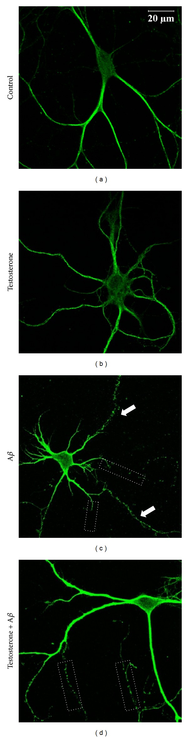
Testosterone reduced neurite shortening following oligomeric Aβ treatment in primary hippocampal neurons. Primary hippocampal neurons were treated with 10 nM testosterone for 1 h, followed by exposure to 5 μM oligomeric Aβ for 24 h. Neurons were stained with MAP-2 antibody. (a) Control, (b) 10 nM testosterone for 25 h, (c) 5 μM Aβ for 24 h, and (d) 10 nM testosterone for 1 h, followed by exposure to 5 μM Aβ for 24 h. Arrows indicate fragmentation of neurites and white dot boxes indicate shortening of neurites.