
Figure 3.
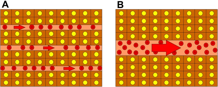
Schematic presentation of the sequential changes to the tumor sinusoidal structure during multistep hepatocarcinogenesis. (A) Well-differentiated hepatocellular carcinoma with a large number of thin sinusoids present; (B) poorly differentiated hepatocellular carcinoma with a relatively small number of large sinusoids present. Note that the total area of the tumor sinusoids is the same as that of (A).