
Figure 4.
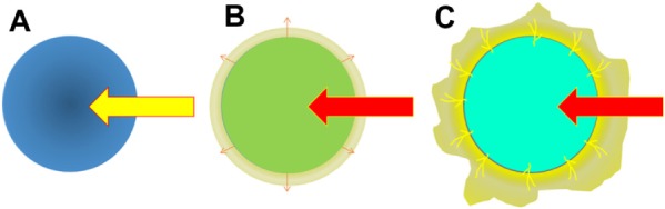
Schematic presentation of the venous drainage of hepatocellular neoplasms during multistep hepatocarcinogenesis.
Notes: (A) Hypovascular dysplastic nodules or early hepatocellular carcinoma. Because venous blood drains out of the lesions via vein-like vessels that connect to the venous system of the surrounding nontumorous tissue, there is no “corona-like enhancement” around the lesion. (B) Hypervascular well-differentiated hepatocellular carcinoma without pseudocapsule. Vein-like vessels decrease in number most likely due to elevated intratumoral pressure caused by increased cellularity. At the periphery of the lesion, the tumor typically shows a “replacing growth pattern”, in which the tumor sinusoids directly face the sinusoids of the surrounding nontumorous hepatocytes. Venous blood from the lesions drains into the surrounding liver tissue via the “trans-sinusoidal pathway”, which can be radiologically observed as a “thin ring-like enhancement” or “thin corona enhancement” on the second phase of CT during hepatic arteriography. An actual case corresponding to this is shown in Figure 5 (A–C). (C) Hypervascular moderately to poorly differentiated hepatocellular carcinoma with pseudocapsular formation. Because of the presence of a pseudocapsule, sinusoids of the tumor and the surrounding liver tissue do not have direct contact in these lesions. Venous blood drains out of the lesions via residual portal venous branches present within the pseudocapsule. These portal venous branches drain into the sinusoids of the surrounding liver tissue, and subsequently, into the venous system, which can be radiologically observed as a typical “corona-like enhancement” on the second phase of CT during hepatic arteriography. An actual case corresponding to this is shown in Figure 5 (D–F).
Abbreviation: CT, computed tomography.