
Figure 6.
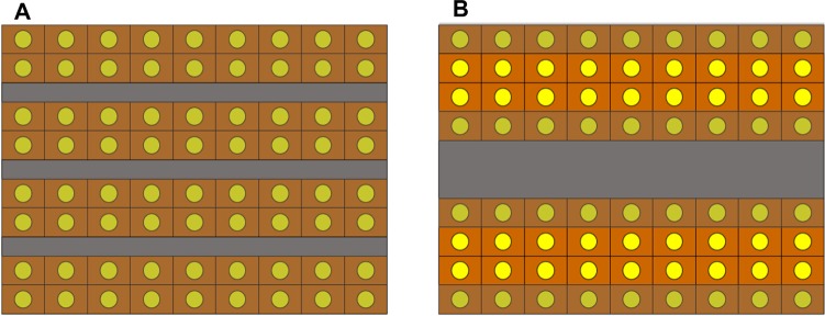
Schematic presentation of the relationship between the distribution of Lipidol emulsion/suspension and histological grades of hepatocellular carcinomas.
Notes: (A) Well or moderately differentiated hepatocellular carcinoma. Because the total contact area of the tumor cells to Lipiodol is large, the chemotherapeutic agent may affect all of the tumor cells. Tumor cells that may be influenced by chemotherapeutic agents are shown in dark color. (B) Poorly differentiated hepatocellular carcinoma. Although the total area of tumor sinusoids is the same as that of (A), the contact areas between Lipiodol and the tumor cells are smaller in (B) compared to (A). Tumor cells that are located in the central part of the thick tumor plates may be less influenced by chemotherapeutic agents (shown in lighter color), whereas those having direct contact with the tumor sinusoids are more likely to be affected by chemotherapeutic agents (shown in darker color).