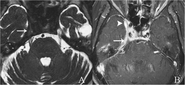
Figure 4.

Axial magnetic resonance imaging at the level of the cavernous sinuses. A. Thin-slice axial T2-weighted images obtained by three-dimensional driven equilibrium sequence revealed edematous right trigeminal ganglion (arrow). B. Postgadolinium-pentetic acid spin echo T1-weighted with fat suppression magnetic resonance imaging of temporal bone. Edematous right trigeminal ganglion within right Meckel’s cave (arrow) surrounded by abnormal prominent enhancement in right Meckel’s cave and cavernous sinus (arrowhead).
