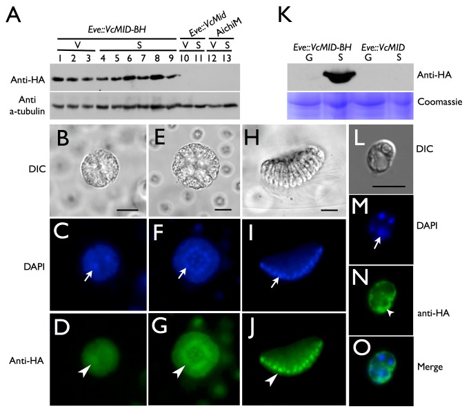
Figure 3. Cell-type restricted expression and sex-regulated nuclear localization of VcMid.
(A) Immunoblot of SDS-PAGE fractionated protein extracts from HA-tagged pseudo-male strain Eve::VcMID-BH (lanes 1–9), untagged pseudo-male strain Eve::VcMid (lanes 10, 11), and wild-type male strain AichiM (lanes 12, 13). Lanes 1–3 contain extracts from vegetative spheroids at adult stage, mid-cleavage stage, and unhatched juvenile stage respectively. Lanes 4–9 contain extracts from spheroids undergoing sexual development with pre-cleavage stage, mid-cleavage stage, unhatched juvenile stage, cleaving androgonidia stage, and mature sexual adult stage, respectively. See also Figure S2B and S2C. Lanes 10 and 12 contain extracts from adult vegetative-stage spheroids. Lanes 11 and 13 contain extracts from mature sexual-stage spheroids. The bands in the upper panel are VcMid-BH protein detected with an anti-HA antibody. The bands in the lower panel come from the same blot re-probed with an anti-tubulin antibody as a loading control. (B–J) DIC (B, E, H) or false-colored IF images of cleaving androgonidia from Eve::VcMID-BH sexual germ cells at the two-cell stage (B–D), sixteen-cell (E–G) stage, and from a mature sperm packet (H–J). IF samples were stained with DAPI shown in blue (C, F, I) or with anti-HA shown in green (D, G, J). Arrows and arrowheads show locations of a representative nucleus from each image. Scale bars = 10 µm. (K) Upper panel, anti-HA immunoblot of SDS-PAGE fractionated protein extracts of purified vegetative gonidia (G) or somatic (S) cells from Eve::VcMID-BH and Eve::VcMID spheroids. Lower panel, Coomassie-stained gel used as a loading control. (L–O) IF detection of VcMid-BH protein from a representative vegetative somatic cell of Eve:: VcMID-BH stained as in (B–J). The arrows in (M, O) show the nucleus, while the chloroplast nucleoids (smaller DAPI-stained spots) are unlabeled. The VcMid-BH signal in (N, O) is excluded from the nucleus as evident in the merged image (O). Scale bar = 7.5 µm.

