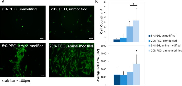
Figure 5.
Panel A shows representative images (scale bars = 100 μm) of each group showing increased adhesion and spreading on modified gels, with the greatest degree of spreading on stiff, modified hydrogels. Panel B shows the results of image analysis of hMSCs seeded onto amine modified and unmodified 5 and 20% w/v PEGDA hydrogels. On both soft and stiff substrates, the cell number (top) was statistically greater on modified gels vs unmodified gels (*, p < 0.01). The average cell area (bottom) on amine modified 20% w/v hydrogels was statistically greater than that of all other groups, indicating cell spreading on the surface of the hydrogel (*, p < 0.05).

