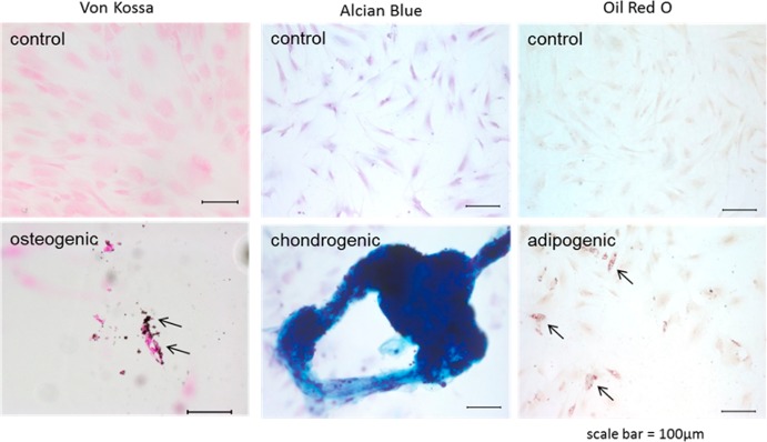
Figure 8.

Histological images showing the results of differentiation on modified hydrogels. Results show that FN modified gels do not induce differentiation without medium induction (top). Images on the bottom panel show the development of calcium deposits which are stained black in color as a result of Von Kossa, the production of a cartilaginous matrix which has stained darkly for the presence of glycosaminoglycans (blue) using Alcian Blue, or the presence of lipid vacuoles which are stained red in color with Oil Red O. All images were taken at 20×, and scale bars are equal to 100 μm.
