
Figure 8.
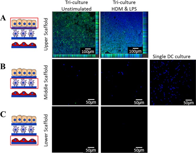
Confocal microscopy images (×10 mag) of DC migration 36 h after allergen stimulation in 3D tricoculture. The upper scaffold contained the Calu-3 epithelial layer (A), a middle layer to which DCs were seeded (B), and a lower scaffold containing MRC-5 (C). DCs were prestained with Hoescht nuclear stain (blue) and Calu-3 cells were poststained with pancytokeratin (green). In single culture, DCs remained on the middle scaffold where they had been inoculated. In the triculture, most DCs migrated from middle scaffold (B) to upper scaffold (A). Upon treatment of the triculture with house dust mite extract (HDM) (10 μg/mL) and lipopolysaccharide (LPS) (100 ng/mL), DCs appeared to primarily migrate to the apical surface of the epithelial layer, whereas in unstimulated samples they appear to be mainly localized in the basal region of the epithelial layer (A). Experiments were performed in duplicates.