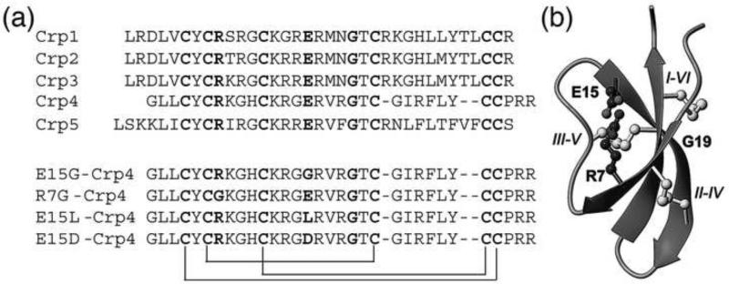
Figure 1.
(a) Sequence comparison of mouse cryptdins. The conserved six cysteines, Gly, and the Arg and Glu involved in the conserved salt-bridge are highlighted in bold and the disulfide connectivities indicated by connecting lines. The sequences of the Crp4 analogues (E15G), (E15L) and (R7G) that have been characterized in this study are shown in the bottom part of the figure. (b) Structural representation of α-defensins. The structure is dominated by the central β-sheet. The side chains of the conserved disulfide array and the salt-bridge are shown in ball-and-stick. The cysteines are labelled with roman numbers I-VI in the order of appearance in the sequence.

