
Fig. 6.
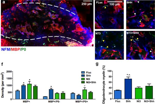
Source of myelination. (a) Widefield view of immunofluorescence of Schwann cell- (NFM+/MBP+/P0+: blue/red/green, respectively) and oligodendrocyte- (NFM+/MBP+/P0−) derived myelin fibers from bridge implants delivering lentivirus encoding SHH. Dashed line indicates border between bridge implant and host tissue. Immunofluorescence at higher magnification from bridges delivering (b) FLuc, (c) SHH, (d) NT3, or (e) NT3+SHH. White arrows show fibers wrapped by Schwann cell-derived myelin and yellow arrows show fibers myelinated by oligodendrocytes. Brightness and contrast were adjusted for clarity. (f) Quantification of total axon numbers in FLuc, NT3, SHH and NT3 and SHH conditions (mean +/− SD). (g) Percentage of axons that were myelinated by oligodendrocytes (mean +/− SD).