
FIGURE 1.
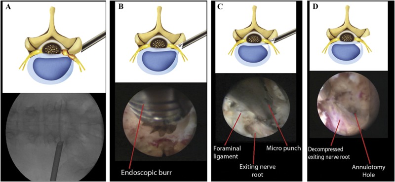
Schematic illustrations and corresponding intraoperative fluoroscopic and endoscopic views of the surgical procedure. A, extraforaminal placement of the working cannula for foraminal decompression. B, foraminal unroofing using an endoscopic burr. C, sophisticated foraminal decompression using various instruments, including punches, forceps, and a laser. Note the exiting nerve root decompression using a micropunch. D, final view of the full-scale foraminal decompression status. Note the decompressed exiting nerve root and annulotomy site.