
FIGURE 3.
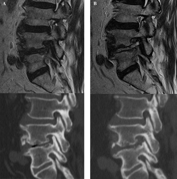
Illustrated case of a 67-year-old male patient. A, preoperative magnetic resonance and computed tomography (CT) images showing severe foraminal stenosis with disc herniation and facet impingement at the left L4-5 level. B, postoperative MR and CT images showing complete foraminal unroofing and visualization of exiting nerve root after endoscopic lumbar foraminotomy.