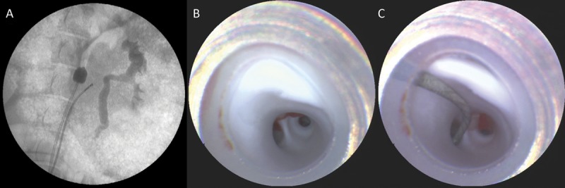
FIGURE 2.

A, fluoroscopic image of the scanning fiber endoscope (SFE) in place in the renal artery with a balloon in the descending aorta for proximal flow control. B, view from the SFE at the end of the guide catheter, showing a smooth white endothelium and the first bifurcation of the renal artery. C, SFE image of renal branch selection using a 0.014-inch microwire.
