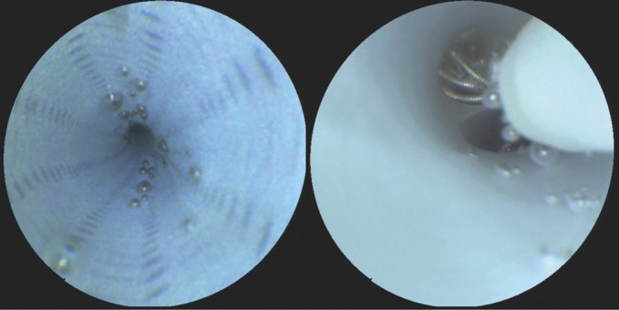
FIGURE 4.

Left, scanning fiber endoscope image demonstrating persistent microbubbles within the lumen of a fastidiously prepared guide catheter that were observed to dislodge randomly with increased saline solution flush rates. Right, image showing the displacement of microbubbles trapped within a flow-diverting stent during device deployment.
