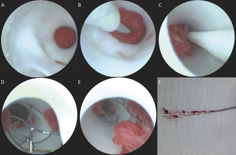
FIGURE 5.

Scanning fiber endoscope images of ischemic stroke thrombectomy from initial lesion inspection (A), microwire crossing (B), initial withdrawal (C), inspection of stent retriever contents in situ (D, E), and ex vivo correlative image of retrieved material (F).
