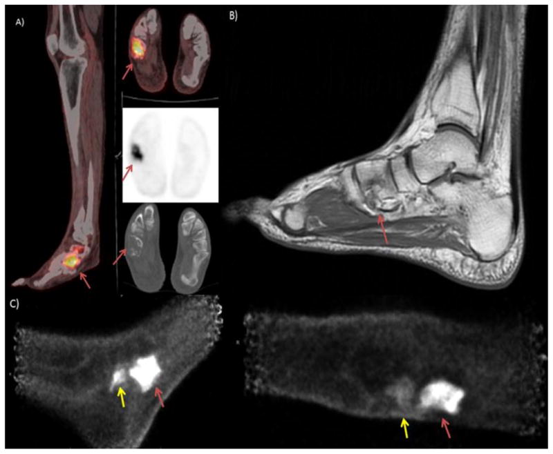
Figure 1.

The patient is a 48 year old man who presented with right foot pain and was found to have a localized cuboidal spindle cell sarcoma which was treated with adjuvant chemotherapy. Disease was moderately refractory to treatment, the PET/CT image shown in image A) demonstrates persistent, intense uptake in a destructive lesion in the right cuboidal bone. Image B) Sagittal MRI with T2 images above and T1 images below show complete marrow replacement in the cuboid on the T2 images with heterogeneous signal in the calcaneus. This was initially read as indeterminate activity in the calcaneus but retrospectively appears suspicious for calcaneal involvement. C) PEM images show viable tumor in the cuboidal bone and increased uptake adjacent to that lesion in the distal calcaneus (yellow arrow) indicating presence of disease extending outside the cuboidal bone.
