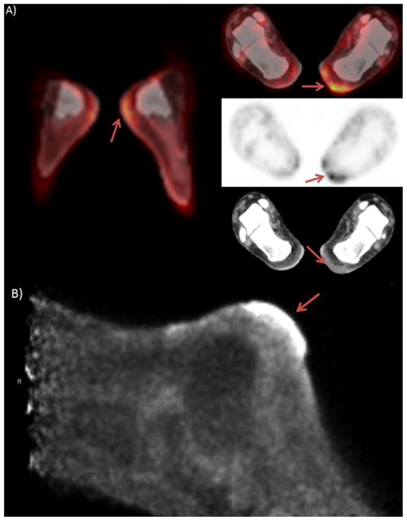
Figure 5.

Patient is a 60 year old female diagnosed with melanoma of the left heel. Patient had surgical excision of a lentiginous melanoma of the left heel five years prior. Patient presented with a left inguinal mass and on PET/CT images A) show mild, diffusely increased uptake of the left heel. PEM images B) show moderate to markedly increased activity of the superficial soft tissues of the left heel with demonstrably superior spatial resolution.
