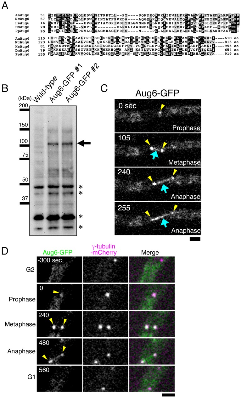
Figure 1. Aug6 is localised at the SPB and the spindle.
(A) Sequence alignment of Aug6 proteins from A. nidulans (An), the red bread mould Neurospora crassa (Nc), Homo sapiens (Hs; also called hDgt6/FAM29A/HAUS6), Drosophila melanogaster (Dm; also called Dgt6), and the moss Physcomitrella patens (Pp). Identical amino acids are boxed, and similar ones are hatched. (B) Detection of Aug6-GFP by immunoblotting. A unique band with the expected molecular weight (arrow) was detected by immunoblotting with the anti-GFP antibody in 2 independent GFP-integrated strains (#1 and #2). Asterisks indicate cross-reactions of the antibody with other proteins. The #2 strain was used throughout this study. (C) Time-lapse imaging of Aug6-GFP during mitosis. Images were acquired every 15 s in a single focal plane. Strong signals were detected at the pole of the spindle (yellow), whereas weak punctate signals were observed along the spindle MT (blue). (D) Time-lapse imaging of Aug6-GFP and γ-tubulin-mCherry. They were co-localised at the SPB during mitosis. See also Movie S1. Bars, 2 µm.

