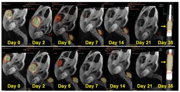
Fig. 2.
Image montages illustrating clearance patterns of I-124 bevacizumab (top) and I-124 ranibizumab (bottom) within the vitreous cavity after pars plana lensectomy in a rabbit model. Note the I-124 accumulation in the thyroid gland. A phantom containing I-124 bevacizumab (top) and I-124 ranibizumab (bottom) in a tuberculin syringe is easily discerned on Day 35 (arrow). Range of acquisition of radioactive emission is 10% to 75%.

