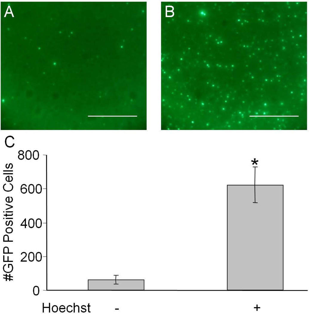
Figure 1.
Human airway epithelia were transduced with 105 vg/cell with AAV5-CMV-eGFP and treated with 0 µM or 10 µM Hoechst-33342 and fluorescent images were taken 7 days later (A and B respectively). Scale bar = 200 µm. The number of GFP positive cells per field of view was quantified and shown in C. (*p<0.01)

