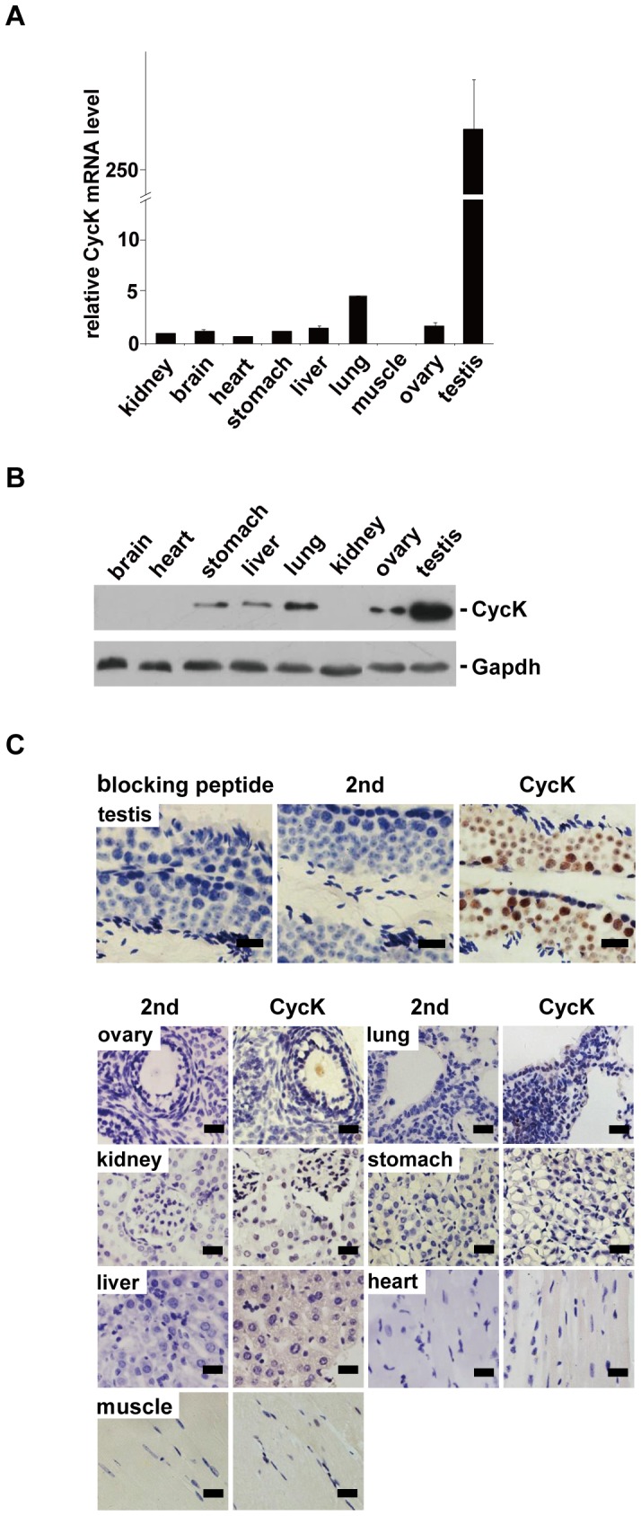
Figure 1. Cyclin K is highly expressed in murine testes.

A) The level of CycK mRNA was determined by qPCR. Expression of CycK was normalized to that of Gapdh in three independent experiments. CycK expression in stomach was arbitrarily assigned to be one, and expression levels in other tissues were normalized to that of stomach. Error bars represent SD. B) Protein blot analysis of CycK expression in different tissues. Total tissue lysates were separated by SDS-PAGE and probed by the indicated antibodies. Representative blot of three independent experiments was shown. C) Tissue sections from 2-month-old mice were stained by IHC with anti-CycK antibodies followed by staining with hematoxylin. Upper panel, detection of CycK in testes. Preincubation with epitope peptides eliminated the signal (left). Secondary antibodies alone did not generate any signal (middle). Cyck expression could be seen in different cell types in seminiferous tubules (right). Lower panel,CycK expression in various tissues. In each case, second antibodies alone (2nd) did not produce detectable signals. Of note, staining was carried out on the same day to allow semi-quantitative comparisons of CycK expression levels. Representative results from four independent experiments were shown. Scale bar: 20 µm.
