
Figure 2. Morphological comparison of three different mouse lines (VM/+, RC/+, RW/+) bearing human HCM causing mutations in the myosin heavy chain gene.
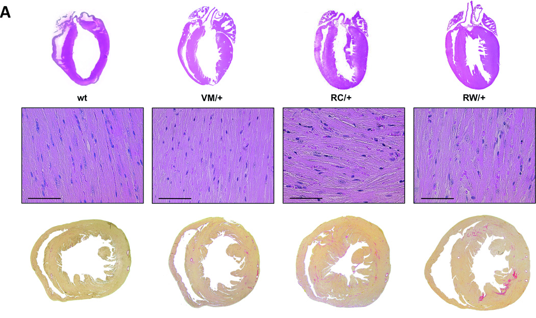
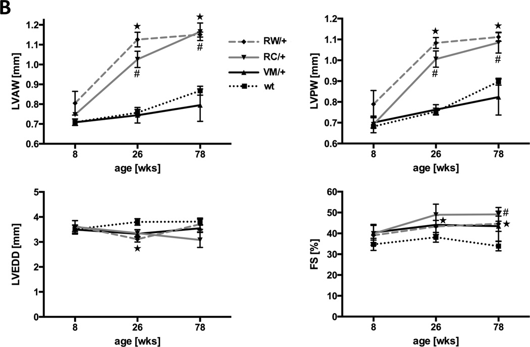
A, Sections through wild type hearts and heterozygous HCM mutants at the age of 26 weeks show concentric hypertrophy, myofiber disarray and interstitial fibrosis only in RC/+ and RW/+ mice. Above, hematoxylin and eosin staining, scale bar, 100 µm. Below, transverse sections, collagen is stained in red by Sirius Red. B, echocardiographic measurements at the age of 8, 26 and 78 weeks, LVAW, left ventricular anterior wall thickness; LVPW, left ventricular posterior wall thickness; LVEDD, left ventricular end diastolic diameter; FS, fractional shortening; n ≥ 3 animals per genotype; *P<0.05, PW/+ vs. wt; #P<0.05, RC/+ vs. wt.