
Figure 4. Characterization of mice homozygous for the human R453C (RC/RC) or R719W (RW/RW) mutation in the α-MHC gene.
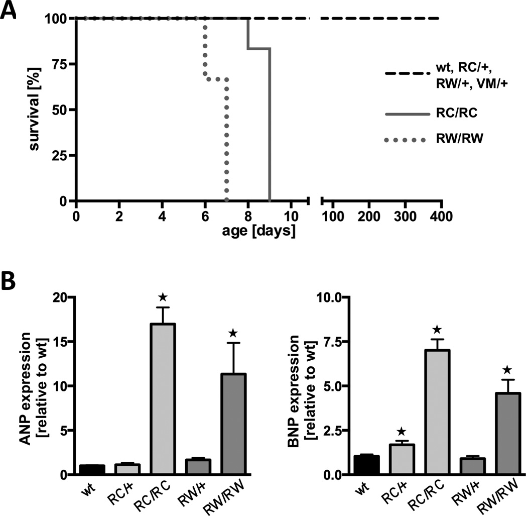
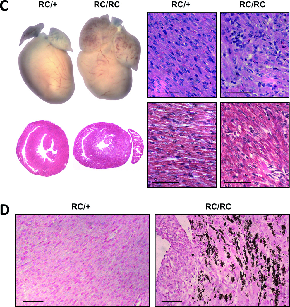
A, survival curves of RC/RC and RW/RW mice compared to heterozygous and wild type (wt) mice; log rank test, P<0.001. B, real time PCR for atrial natriuretic peptide (ANP) and brain natriuretic peptide (BNP) in 7-days-old ventricles. C and D, macroscopic and microscopic view of heterozygous and homozygous RC mutants at age 7 days; *P<0.05 vs. wild type. C, left, whole hearts and transverse sections (note the enlarged atria of RC/RC hearts); right, heart sections stained with hematoxylin and eosin (above) and with Masson’s trichrome (below); scale bar, 50 µm. D, vonKossa staining of left ventricular tissue indicates areas of calcification (brown staining) only in homozygous hearts (right panel); scale bar, 50 µm. Equivalent findings were made in RW/RW hearts.