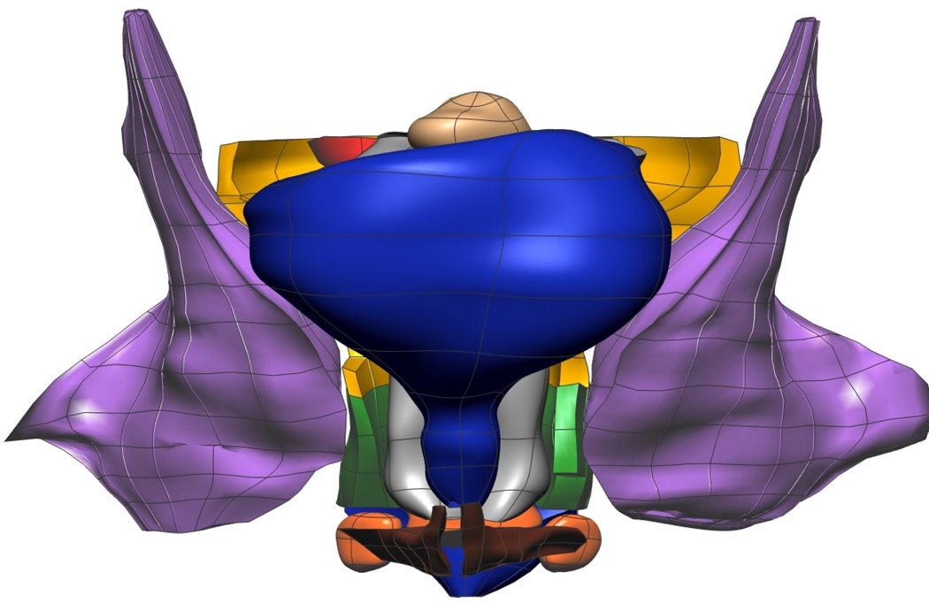
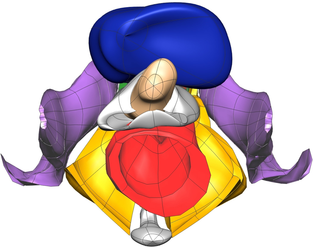
Fig. 10. Subject-specific MRI Mesh Views.
Final fitted meshes from the MRI data set. Shown are (a) anterior, (b) posterior, (c) cephalad and (d) caudad views showing 12 of the 13 components – PR (green), LA (gold), EAS (blue), IAS (beige), rectum (red), TP (orange), PB (orange), coccyx (silver), uterus (beige), vagina (silver), OI (purple) and bulbospongiosus (brown). For clarity the lumen has not been included.


