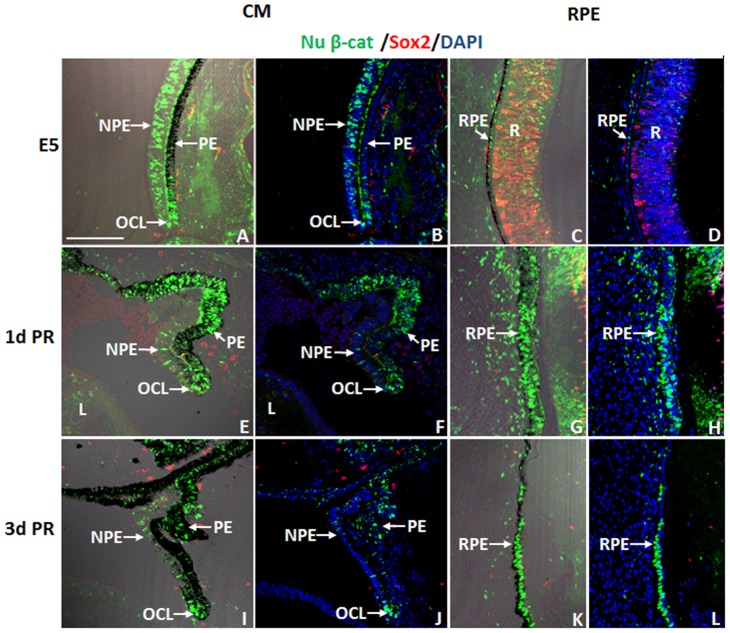
Figure 4. Dynamic changes of nuclear β-catenin in the chick eye after injury.
(A–L) Presence of nuclear β-catenin (Nu β-cat) and Sox2 in the CM (A, B, E, F, I, J) and RPE (C, D, G, H, K, L) at E5 (A–D), at 1 d PR (E–H) and at 3 d PR (I–L). Panels A, C, E, G, I, K include DIC overlay and are equivalent to B, D, F, H, J, and L respectively. NPE: non-pigmented epithelium; PE: pigmented epithelium; OCL: optic cup lip; RPE: retinal pigmented epithelium; R: retina. DAPI stains the nuclei of the cells in B, D, F, H, J and L. Scale bar in (A) represents 100 µm and applies to all panels.

