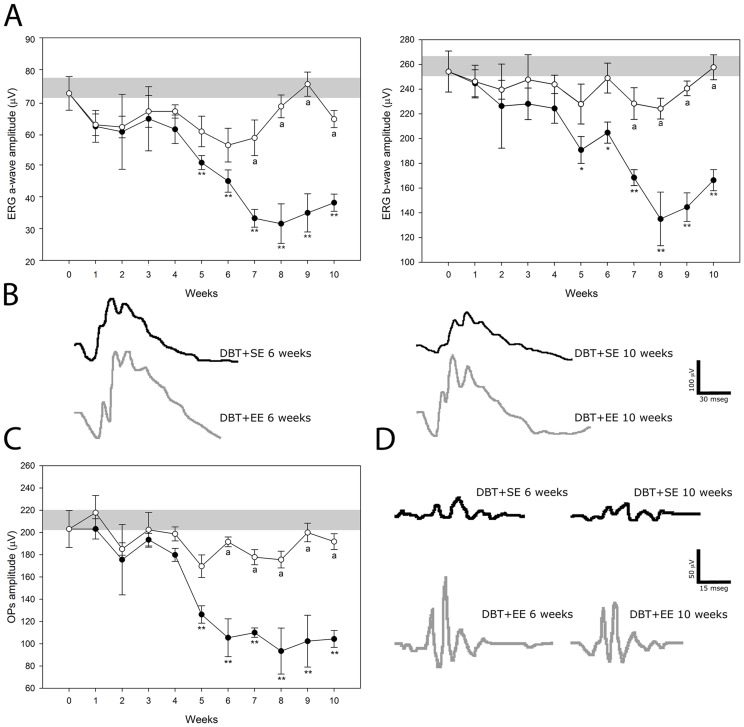
Figure 2. Effect of EE housing on retinal dysfunction induced by experimental diabetes.
ERGs (A) and OPs (C) were weekly registered after the induction of diabetes. Experimental diabetes induced a significant decrease in ERG a- and b-wave amplitude in eyes from animals housed in SE (black circles), as compared with age-matched control animals (grey bar), after 5 weeks of STZ injection. In diabetic eyes from animals housed in EE (white circles), a significant prevention of these alterations was observed. Panel B shows representative scotopic ERG traces from control and diabetic animals housed in SE (DBT+SE) or EE (DBT+EE) assessed at 6 and 10 weeks after diabetes induction. A significant decrease in the sum of OP amplitude (C) was observed after 5 weeks of diabetes onset in eyes from animals housed in SE, while EE housing prevented the effect of diabetes on this parameter. Panel D shows representative OP traces from control and diabetic animals housed in SE or EE and assessed at 6 and 10 weeks after induction of diabetes. Data are the mean ± SEM (n = 10–12 animals per group); *p<0.05, **p<0.01 versus age-matched control; a: p<0.01 versus diabetic animals in SE, by Tukey's test.

