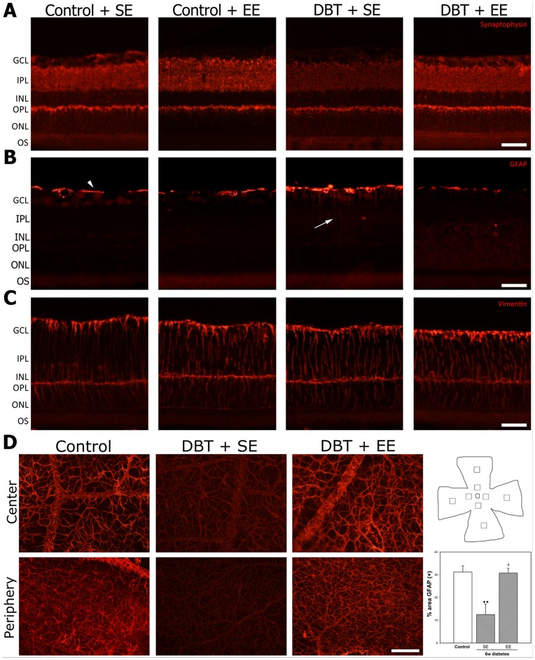
Figure 4. Effect of EE housing on retinal synaptophysin- and Müller cell and astrocyte GFAP-immunoreactivity.
Panel A: Representative photomicrographs of retinal sections from a vehicle injected animal housed in SE or EE, and a diabetic animal housed in SE or EE. At 6 weeks of experimental diabetes, a decrease in IPL synaptophysin levels was observed in SE-housed animals, whereas EE housing prevented the decrease in synaptophysin immunoreactivity in the IPL from diabetic rats. No evident differences in synaptophysin levels were observed in the ONL among groups. In non-diabetic animals, EE did not affect synaptophysin immunoreactivity. Shown are photographs representative of four eyes per group. Panel B: In SE-housed diabetic animals, a slight increase in Müller cell process GFAP levels (arrow) was observed which was not evident in EE-housed diabetic animals Panel C: no changes in vimentin-immunoreactivity were observed among groups. Panel D: Assessment of astrocyte GFAP-immunoreactivity in flat-mounted retinas after 6 weeks of diabetes induction. In control retinas, astrocytes were intensely immunoreactive for GFAP in the central and peripheral regions. The intensity of GFAP-immunoreactivity was significantly reduced in eyes from diabetic animals housed in SE, both in the peripapillary and the peripheral regions. In eyes from diabetic animals housed in EE, a prevention of the decrease in GFAP-immunoreactivity induced by diabetes was observed both in central and peripheral regions. Right panel: Schematic diagram of a flat-mounted retina showing all the regions analyzed. Assessment of the GFAP immunoreactivity area. Data are the mean ± SEM (n = 5 eyes per group); **p<0.01, versus age-matched controls; a: p<0.01, versus diabetic animals housed in SE, by Tukey's test. Scale bar: 50 µm for panel A, B and C, and scale bar: 100 µm for panel D.

