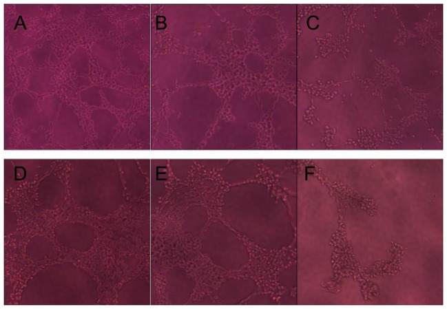
Figure 4. The effect of HLC-080 on EA.hy926 vascular tube formation.
EA.hy926 were seeded on matrigel in 96-well plates at 3×10⁴ cells per well. The cells were mixed with different concentrations of HLC-080 and photographed at various time points, as described in the methods. (A) Control at 6 h showing EA.hy926 without additional stimuli. (B) HLC-080 1.0 µM added to EA.hy926, showing vascular tubes at 6 h. (C) HLC-080 5.0 µM added to EA.hy926, showing vascular tubes at 6 h. (D) Control at 24 h showing EA.hy926 without additional stimuli. (E) HLC-080 1.0 µM added to EA.hy926 formed only a small number of short, incomplete tubes at 24 h (F) HLC-080 5.0 µM added to EA.hy926, exhibited more significant effects at 24 h.

