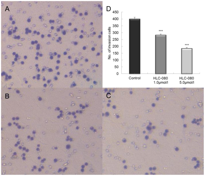
Figure 5. The effect of HLC-080 on cell invasion in HT-29 cells.
Cells were seeded in a transwell chamber and allowed to migrate across the chamber toward cell-specific conditioned medium for 24 h. Photomicrographies of stained migrating cells were taken under brightfield illumination. Representative images are shown for HT-29 control (A) and HT-29 disposed by HLC-080 1.0, 5.0 µM (B, C). Quantification of the invasion is expressed as the number of invasive cells in five random microscopic fields per well (mean ± SD; ***p<0.001 versus control group cells) (D). Results were obtained from three separate experiments.

