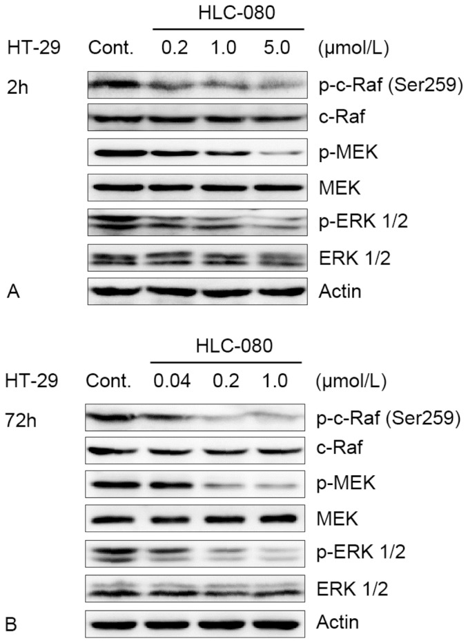
Figure 6. The effect of inhibition of Raf/MEK/ERK pathway in HT-29 cells in vitro.

(A) HT-29 cells were incubated with increasing concentrations of HLC-080 for 2 h. (B) HT-29 cells were incubated with increasing concentrations of HLC-080 for 72 h. Whole-cell lysates were subjected to western blot analysis. For western blot analysis, samples were transferred to a polyvinyldine diflouride membrane by semi-wet electrophoresis and incubated with indicated primary antibody p-c-Raf (Ser259), c-Raf, p-MEK1/2 (Ser217/221), MEK1/2, p-ERK1/2 (Thr202/Tyr204), ERK1/2). Actin was used as loading and transfer control. The experiment was repeated twice and similar results were obtained.
