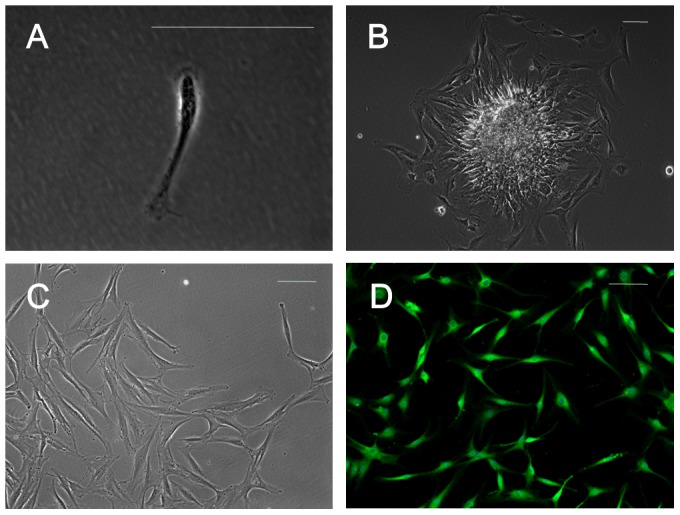
Figure 1. SFMSCs obtained from TMD patients.

(A) Dissociated single adherent cells were observed after culture of synovial fluid samples for 48(B) Cells were observed growing from small tissue masses after culture of synovial fluid samples for 5 days. (C) Microscopic image showing the typical morphology of synovial fluid-derived cells. (D) Immunofluorescent staining of synovial fluid-derived cells demonstrated positive expression of VCAM-1. Scale bars = 100 µm.
