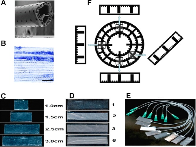
Figure 1.

(A) Scanning electron microscope image of the tubular PCL Swiss-roll construct. (B) Astrocyte distribution within the tubular construct after 2 weeks of culture. The Swiss-roll was unrolled for imaging. Scale bar = 2 mm. (C) Mini-chambers with defined width (1 cm) and different lengths (1.0, 1.5, 2.5, 3.0 cm). (D) Mini-chambers (1 cm wide, 3.0 cm long) with 1, 2, 3, and 6 layers of porous lids. (E) The appearance of the mini-chambers (1 cm wide, 2.5 cm long) with connected silicone tubes as used for time-lapse experiments. (F) Schematic diagram of the cross-sections of a Swiss-roll and mini-chambers rotated accordingly, illustrating how these simulate various positions within the Swiss-roll.
