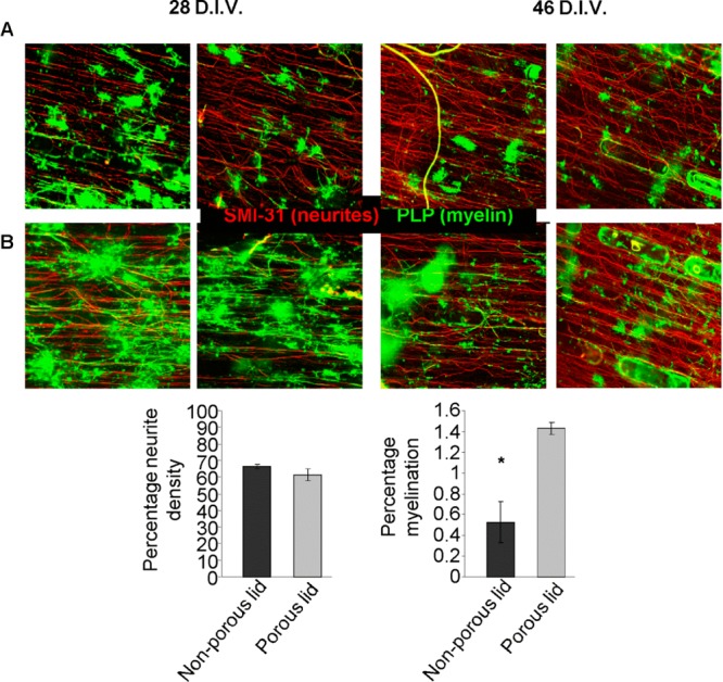
Figure 4.

(A,B) Representative images of myelinating cultures within a 0.5 cm long mini-chamber at both 28 D.I.V. and 46 D.I.V. (A) with a nonporous lid and (B) with a porous lid. All images were labeled for SMI-31 (red) and PLP (green). Graphs show neurite density (C) and percentage of myelination (D) for the myelinating cultures at 28 D.I.V. n = 3. Scale bar: 100 μm.
