
Figure 3.
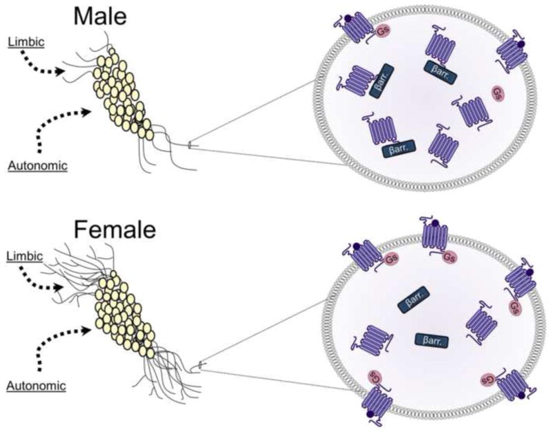
Schematic depicting sex differences in the LC–arousal system in rodents. The images on the left depict LC neurons with dendrites that are longer and more complex in females (bottom left panel) compared to males (top left panel). The longer dendrites of females are more likely to receive limbic afferents that terminate in peri-LC region. However, males and females would likely receive comparable amounts of input from autonomic regions that synapse near the cell bodies. The images on the right depict magnified views of LC dendrites to illustrate sex differences in the CRF1 receptor coupling and trafficking. The top right panel shows how CRF1 receptors (purple) of males associate with βarrestin2 (βarr.) and internalize following stressor exposure. In contrast, the bottom right panel shows how CRF1 receptor of females couple to Gs and traffic to the plasma membrane following stressor exposure.