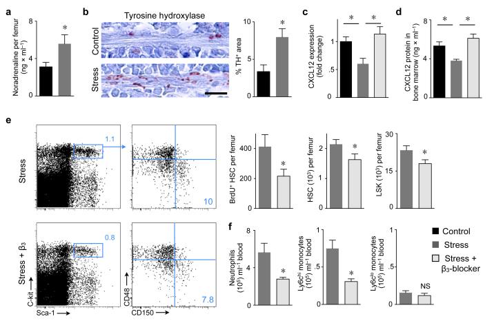
Figure 3. Stress–induced sympathetic nervous signaling regulates proliferation of bone marrow HSC via CXCL12.
a, Noradrenaline ELISA after 3 weeks of stress (n = 8 per group, Student’s t–test). b, Immunoreactive staining for tyrosine hydroxylase (TH) in bone marrow. Scale bar indicates 10 μm. Bar graph shows TH–positive area (n = 5 mice per group, Mann–Whitney test). c, CXCL12 mRNA in bone marrow (n = 10 per group, one–way ANOVA). d, CXCL12 protein in bone marrow (n = 7 per group, one–way ANOVA). e, Dot plots and quantification of LSK and HSC (n = 5 per group, Mann–Whitney test). f, Effects of β3 adrenoreceptor blocker on blood leukocytes (n = 5 per group, Mann–Whitney test). Mean ± s.e.m., * P < 0.05.

