Figure 4.
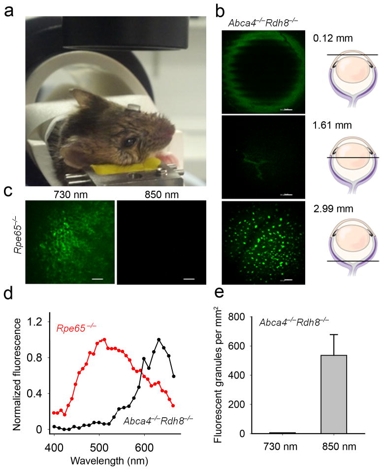
Set-up for two-photon RPE imaging in living mice. (a) During imaging a contact lens covers mouse eye facing the objective. (b) Representative images of a pigmented 7-week-old Abca4−/−Rdh8−/− mouse eye obtained in vivo with 850 nm excitation 14 days after exposure to bright light, at different depths along Z-axis; a 120 μm section through the cornea, a 1608 μm section showing lens sutures, and a 2987 μm section revealing fluorescent granules in the RPE. (c) Images of the RPE in live albino 7-week-old Rpe65−/− mice obtained with 730 nm and 850 nm excitation. (d) Fluorescence emission spectra from RPE of 7-week-old Abca4−/−Rdh8−/− mice obtained with 850 nm and 7-week-old Rpe65−/− mice obtained with 730 nm excitation light in vivo. (e) Quantification of fluorescent granules. Error bars indicate S.D., n = 3.

