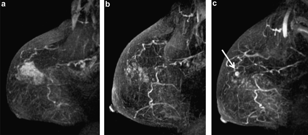
Fig. 1.
58 year old woman with high grade DCIS (patient 12). (a) Baseline MRI shows extensive abnormal clumped ductal enhancement in the upper breast. (b) Breast MRI at 19.3 months on aromatase inhibitor therapy demonstrates improved appearance of abnormal enhancement in the right breast. (c) Breast MRI at 25.5 months since diagnosis demonstrated continual improvement in clumped ductal enhancement but a new 6 mm mass enhancement (white arrow) representing 8 mm of Grade 3 ER−/PR−/Her2neu + IDC at surgery. (Sagittal T1 post gadolinium subtracted maximum intensity projections).

