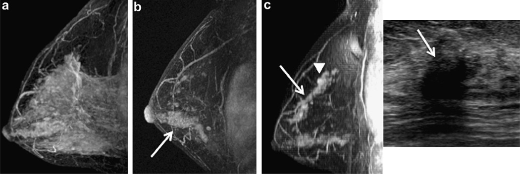
Fig. 2.
47 year old woman with intermediate grade DCIS who discontinued endocrine therapy after 13 months (patient 5). (a) Baseline MRI revealed extensive background enhancement which was dramatically reduced at 3 month follow up where extensive clumped ductal enhancement is seen in the lower breast (arrow) and scattered clumped ductal enhancement is seen in the upper breast (b). (c) 65.5 months from diagnosis, breast MRI showed increasing clumped ductal (arrow, left image) and mass-like (arrowhead, left image) enhancement in the upper breast with a corresponding suspicious hypoechoic mass seen at ultrasound (arrow, right image). Mastectomy revealed multifocal grade 2 IDC (ER+/weakly PR+/Her2neu−) and low to intermediate grade DCIS. (Sagittal T1 post gadolinium subtracted maximum intensity projections (MIP) and targeted ultrasound).

