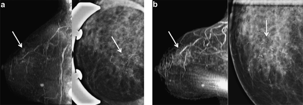
Fig. 3.
55 year old woman with intermediate grade DCIS (patient 10). (a) Baseline MRI with upper inner clumped ductal enhancement (arrow, left image) and pleomorphic microcalcifications at mammography (arrow, right image). (b) 14.5 months after diagnosis breast MRI shows increasing clumped ductal enhancement in a segmental distribution (arrow, left image) and increasing pleomorphic microcalcifications at mammography (arrow right image). Lumpectomy specimen revealed 6 cm of high grade weakly ER+/PR+ and Her2neu + DCIS. (Sagittal T1 post gadolinium subtracted maximum intensity projections (MIP) and CC spot compression magnification mammography).

