Figure 2.
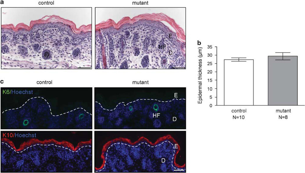
Lack of skin abnormalities in newborn Fst mutant mice. (a) Masson trichrome-stained histological sections of newborn control and Fst mutant skin are shown. (b) Morphometric analysis of tail skin revealed no difference in epidermal thickness. N =number of mice. Results shown are mean ± s.e.m. (c) Expression of keratin 6 (K6; first panel, green) and keratin 10 (K10; second panel, red) is similar in control and mutant mice. Dotted line indicates epidermal–dermal border. (a, c) Bar = 50µm. E: epidermis; D: dermis; HF: hair follicle.

