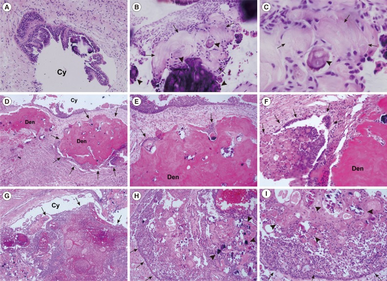
Fig. 2.
Calcifying odontogenic cyst (COC) and calcifying cystic odontogenic tumor (CCOT) photomicrographs. (A-C) COC, cystic epithelium is keratinized and produces irregular calcifications (arrowheads) and aberrantly keratinized ghost cells (arrows). (D-I) CCOT. (D-F) Odontoma-associated CCOT, proliferating tumor mass (arrows) containing dysplastic dentinoid materials (Den). (G-I) Ameloblastomatous proliferating CCOT, infiltratively proliferating tumor cells (arrows), accompanying multiple ghost cell calcifications (arrowheads). Cy, cyst space (Fig. 2D-I; Courtesy of Professor Kyung-Ja Cho, Department of Pathology, University of Ulsan College of Medicine, Korea).

