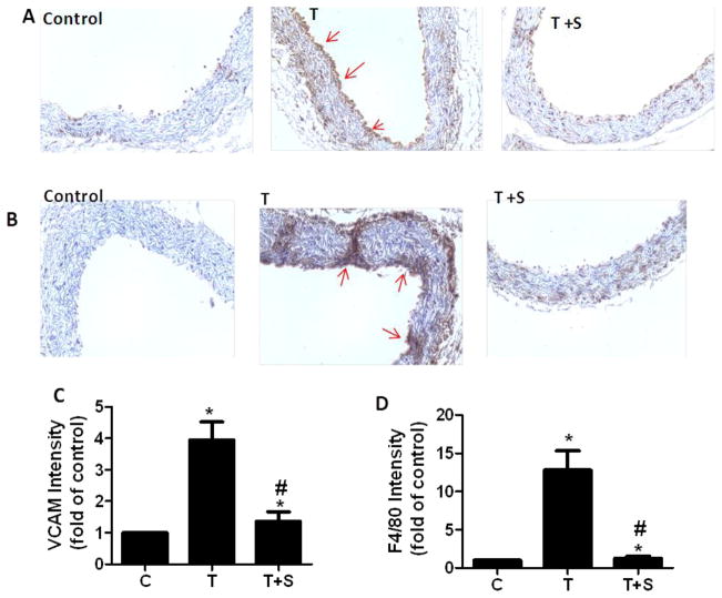
Fig 5. Immunohistochemical staining for adhesion molecule VCAM-1 and F4/80-positive monocytes-derived macrophages in aortic cross-sections.
Representative photomicrographs of immunohistochemical staining for F4/80-positive monocytes-derived macrophages (A) and VCAM-1 (B). Quantitative analysis of VCAM-1 (C) and F4/80 (D). Arrows indicate typical positive-stained regions and original magnification is 40X. T, TNF-α; T +S, TNF-α + sulforaphane. Data are expressed as mean ± SEM, n=5, *, p<0.05 vs. control; #, p<0.05 vs. TNF-α alone-treated mice.

