FIG. 3.

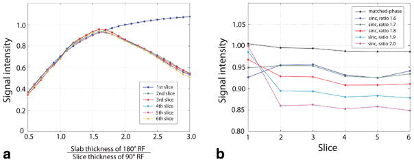
a: Signal intensity of each slice measured in multislice imaging using sinc-based 90°–180° RF pulses with 6-skip-2 slice configuration. For each slice, signal intensity was averaged and normalized by the signal intensity measured in single-slice imaging. For all slices except for the first slice, the signal intensity decreased as the 180° RF slab thickness became larger than 1.6 times the 90° RF slice thickness because of more magnetization perturbation from previous slices. b: Normalized signal intensity in each slice acquired using sinc-based RF with slab thickness ratio 1.6–2.0 and matched-phase RF. [Color figure can be viewed in the online issue, which is available at wileyonlinelibrary.com.]
