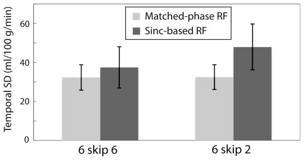
FIG. 7.

Temporal SD of CBF time series measured in normal volunteers using VSASL. Error bars correspond to the ± 1 SD across five subjects. When slice spacing was reduced from 6 to 2 mm, temporal SD increased from 37 mL/100 g/min to 48 mL/100 g/min using sinc-based RF pulses but was preserved at 32 mL/100 g/ min using matched-phase RF. Larger crosstalk is suspected to generate higher temporal SD in the presence of brain motion during image acquisition.
