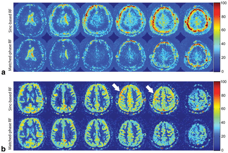
FIG. 8.
Temporal SD maps (a) and corresponding average CBF maps (b) acquired in a healthy volunteer using VSASL with sinc-based RF and matched-phase RF (in mL/100 g/min). All images were acquired with 6-mm slice thickness and 2-mm slice spacing. Using matched-phase RF, temporal SD of CBF of all slices was reduced in slice 2–6, and the hyperintensity in CBF maps (arrows) was reduced as a result of improved ASL signal stability.

