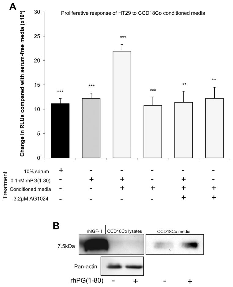
Figure 4.
(A) Increase in RLUs (indicative of proliferation) compared with serum-free media in HT29 cells 18 hours after treatment with 10% serum, 0.1 nmol/ L rhPG(1–80), CCD18Co-conditioned media 18 hours after rhPG(1–80) treatment, CCD 18Co-conditioned media alone, and CCD18Co-con-ditioned media 18 hours after treatment with rhPG(1–80) in the presence of AG1024 or CCD18Co-conditioned media in the presence of AG1024. (B) Western blot showing mature IGF-II expression in CCD18Co cells and secretion by CCD18Co cells into the media with or without rhPG(1–80).**P < .01 and ***P < .001 compared with serum-free media by Student t test.

