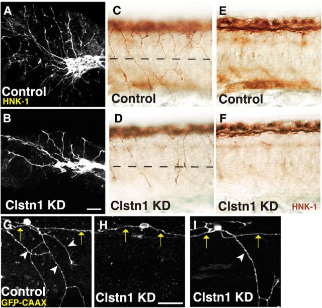
Figure 2.
Clstn-1 knockdown reduces branches in peripheral sensory axons. A, B, Lateral views of trigeminal ganglia labeled with anti-HNK-1 in uninjected (A) and Clstn-1 knockdown (KD) (B) embryos showing reduced branching of trigeminal axons. C, D, Lateral views of RB neurons labeled with anti-HNK-1 showing a reduction in peripheral axons in Clstn-1 KD (D) versus control MO-injected embryos (C). E, F, The focal plane of central axons in the same embryos showing no effect on the central fascicle. G–I, Single-labeled RB cells expressing GFP-CAAX show a reduction of peripheral outgrowth (H) and peripheral branching (I) in Clstn-1 KD embryos. Arrows indicate central axons; arrowheads indicate peripheral axons. Scale bars, 40 μm.

